Osteochondrosis

What is Osteochondrosis?
Osteochondrosis is a common condition that affects the joints of young, rapidly growing dogs. The surface of the joint (the articular cartilage) fails to convert into bone in specific locations. This results in areas of thickened cartilage. These areas are weak and cause the thickened cartilage to detach from the surrounding normal cartilage and form a flap.
This process is called osteochondritis dissecans (or OCD). The flap of abnormal cartilage may detach from the surface of the joint and form what is termed a ‘joint mouse’.
Osteochondrosis causes the development of secondary osteoarthritis. Osteochondrosis primarily affects the shoulder, elbow, knee (stifle) and ankle (hock) joints. The condition frequently affects both left and right joints (termed ‘bilateral’).
What are the symptoms of Osteochondrosis?
Affected dogs may show signs of:
- Lameness
- Stiffness (especially post exercise)
- Lethargy
- Exercise reluctance
- Preference to lie rather than sit or stand
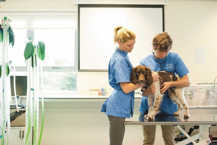
How is Osteochondrosis diagnosed?
Examination may reveal muscle wastage. Manipulation of the affected joint(s) may cause pain. Swelling and restriction in range of movement may be evident.
X-rays are the most common method of diagnosing osteochondrosis. They enable the presence and severity of secondary osteoarthritis to be assessed. A more sensitive way of diagnosing the condition in this joint is by placing a small camera in the joint – this is called arthroscopic examination.
How is a Osteochondrosis treated?
Shoulder Osteochondrosis
Surgery is generally indicated to remove the fragment of loose cartilage. This can be done arthroscopically or via a direct surgical approach. Following removal of the flap of cartilage the defect heals with an inferior type of cartilage called fibrocartilage.
Conservative management of shoulder osteochondrosis is generally not recommended as pain and lameness may persist as long as the cartilage flap remains attached. The results of this approach are uncontrollable and unpredictable.
The majority of dogs with shoulder osteochondrosis recover very well following surgery. Lameness usually resolves despite the development of osteoarthritis. Occasionally stiffness or lameness after vigorous exercise will be evident.
Stifle Osteochondrosis
Surgery is generally advised to remove the fragment of loose cartilage. Following removal of the flap of cartilage the defect heals with an inferior type of cartilage called fibrocartilage. Conservative management is occasionally indicated, especially in older dogs.
Some dogs do quite well following stifle osteochondrosis surgery and others remain lame. It is not possible to predict the outcome in individual cases.
Hock Osteochondrosis
When the condition is diagnosed at a young age, for example six months, surgery is generally recommended to remove the fragment of loose cartilage. Following removal the defect heals with an inferior type of cartilage called fibrocartilage. In selected cases reattachment of loose areas of bone and cartilage can be attempted. If successful this can give a good outcome, although complications can be seen. Conservative management may be appropriate in older dogs that have established osteoarthritis.
Some dogs with hock osteochondrosis develop severe secondary osteoarthritis that results in permanent pain and lameness. If the response to conservative management (weight regulation, exercise restriction, painkillers) is unsatisfactory, salvage surgery may be required. This involves fusing the joint (arthrodesis). A bone graft is packed into the joint following removal of cartilage and the joint is stabilised with a plate and screws.
The outlook with hock osteochondrosis is quite guarded, with many dogs having some degree of persistent stiffness and lameness.
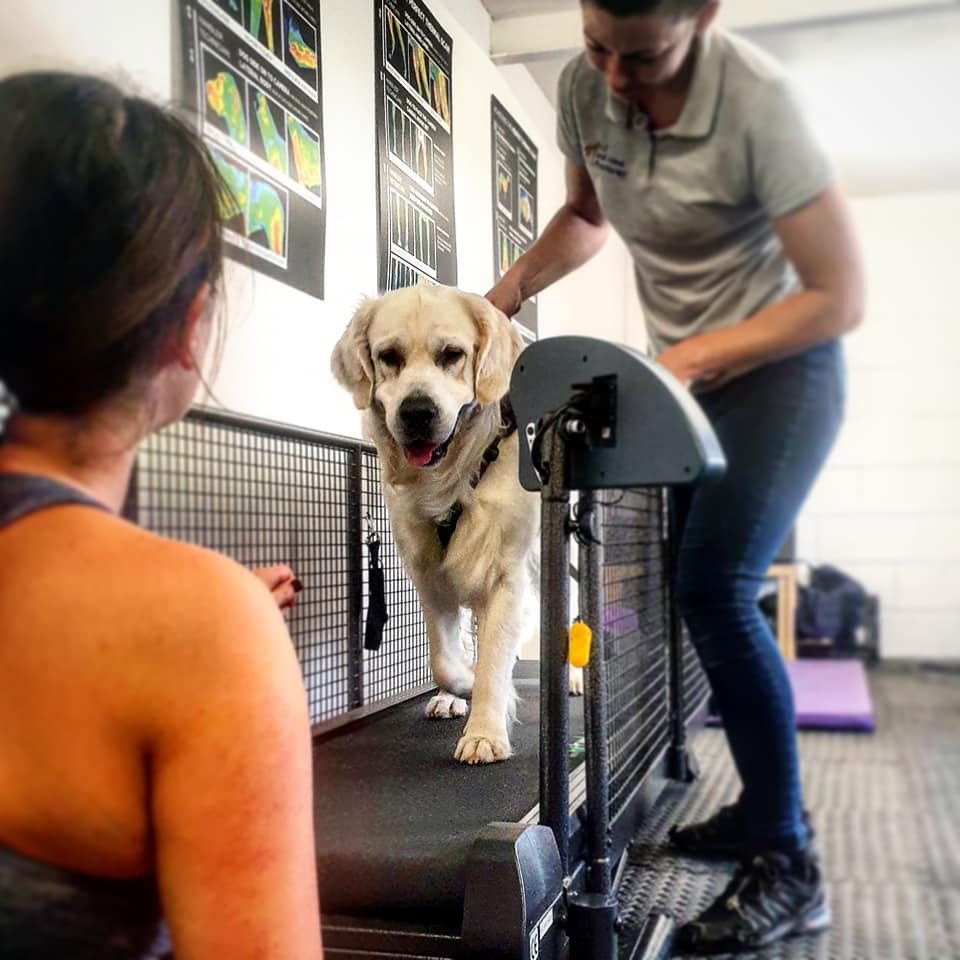
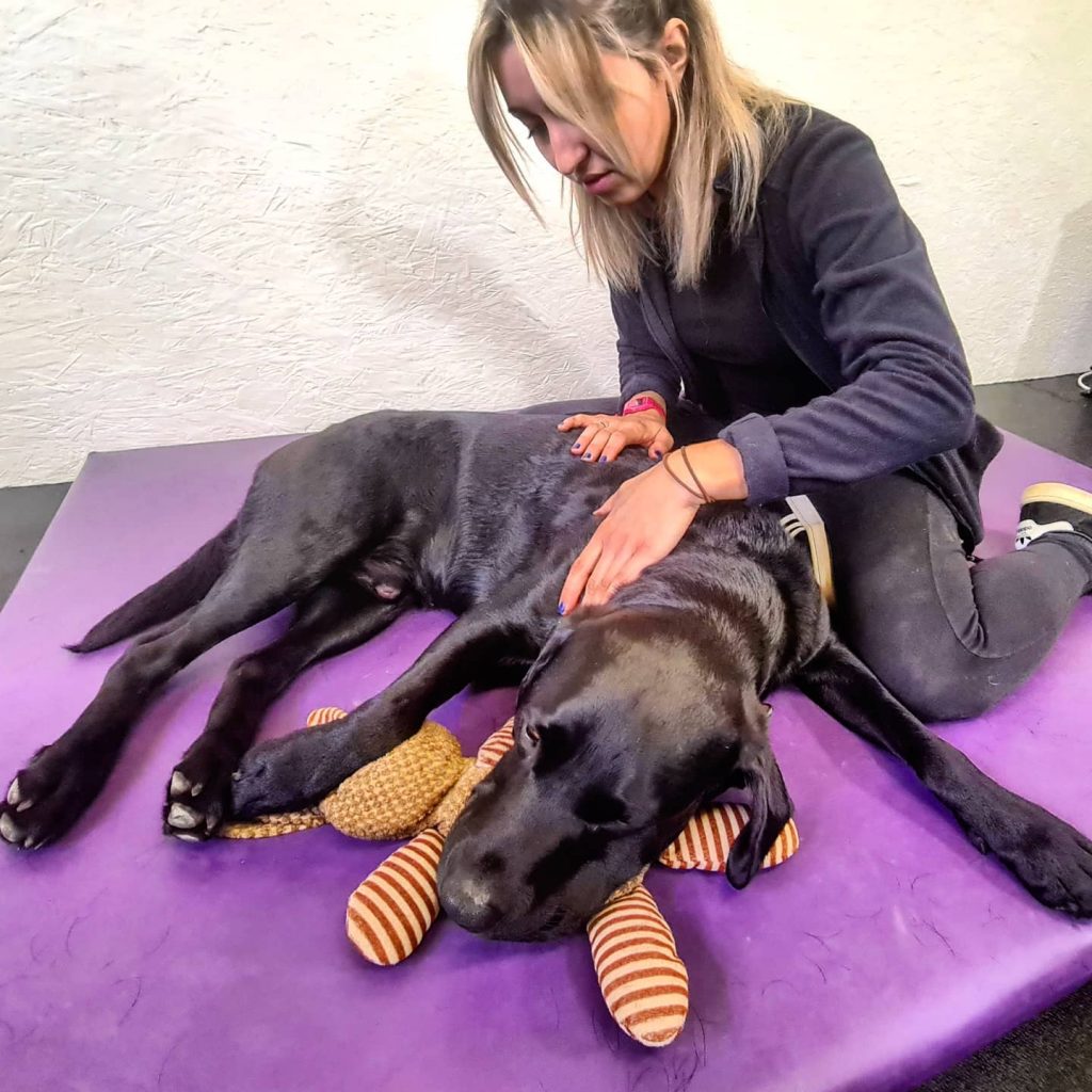
Orthopaedic rehabilitation
Rehabilitation is a process which aims to maximise patient mobility and wellbeing, returning them to their usual way of life following illness, injury or surgery. We restore pets to normal function (or as close as is possible), efficiently and safely using a wide variety of physiotherapeutic techniques.
Injury and even surgery can disrupt the body’s equilibrium in all sorts of direct and indirect ways. Even a pet’s own protective responses such as the inflammatory process can overwhelm and inhibit healing so one objective of rehabilitation is to reduce this level of inflammation. During rehabilitation, we also aim to boost the circulatory system, improve muscle function, increase range of motion within joints, and stimulate innate pain-relieving mechanisms.
With a committed and planned rehabilitation programme, pets can recover more quickly, realise better outcomes and avoid much pain and discomfort.
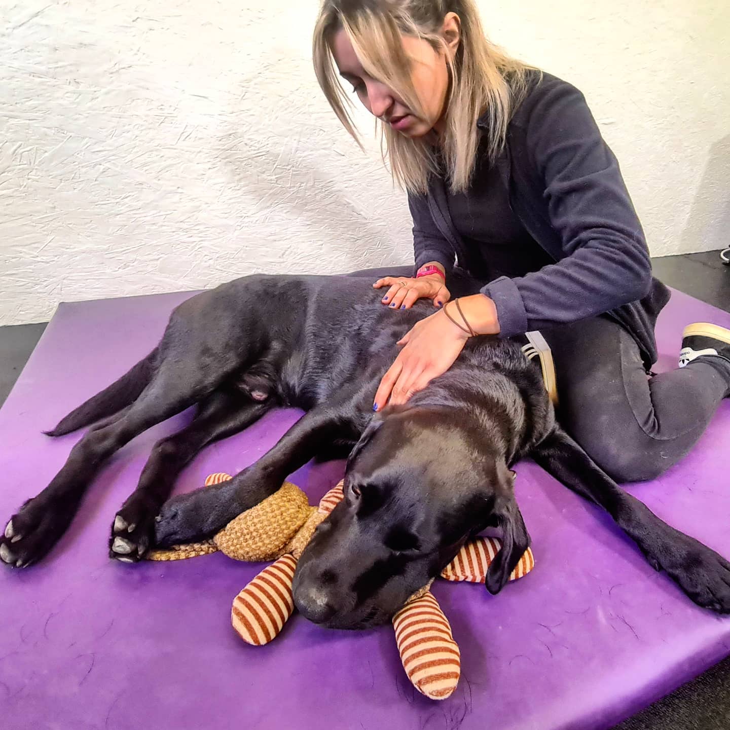
The best rehabilitation programmes consider the whole pet, not just the area of injury; we target and improve multiple systems throughout the body without forgetting the invaluable healing effects of boosting mental wellbeing too. From the wound healing properties of laser treatment, and the muscle strengthening of hydrotherapy, to the circulation boosting effects of massage, we will devise a rehabilitation programme to match a pet’s specific requirements.
Ready to get some help?
Our friendly and skilled physiotherapists are ready to help you and your dog with their rehabilitation.
More conditions
The content on this page is for advice and information only and does not represent veterinary guidance or direction. Please always consult a veterinary surgeon if you are worries about your dog.