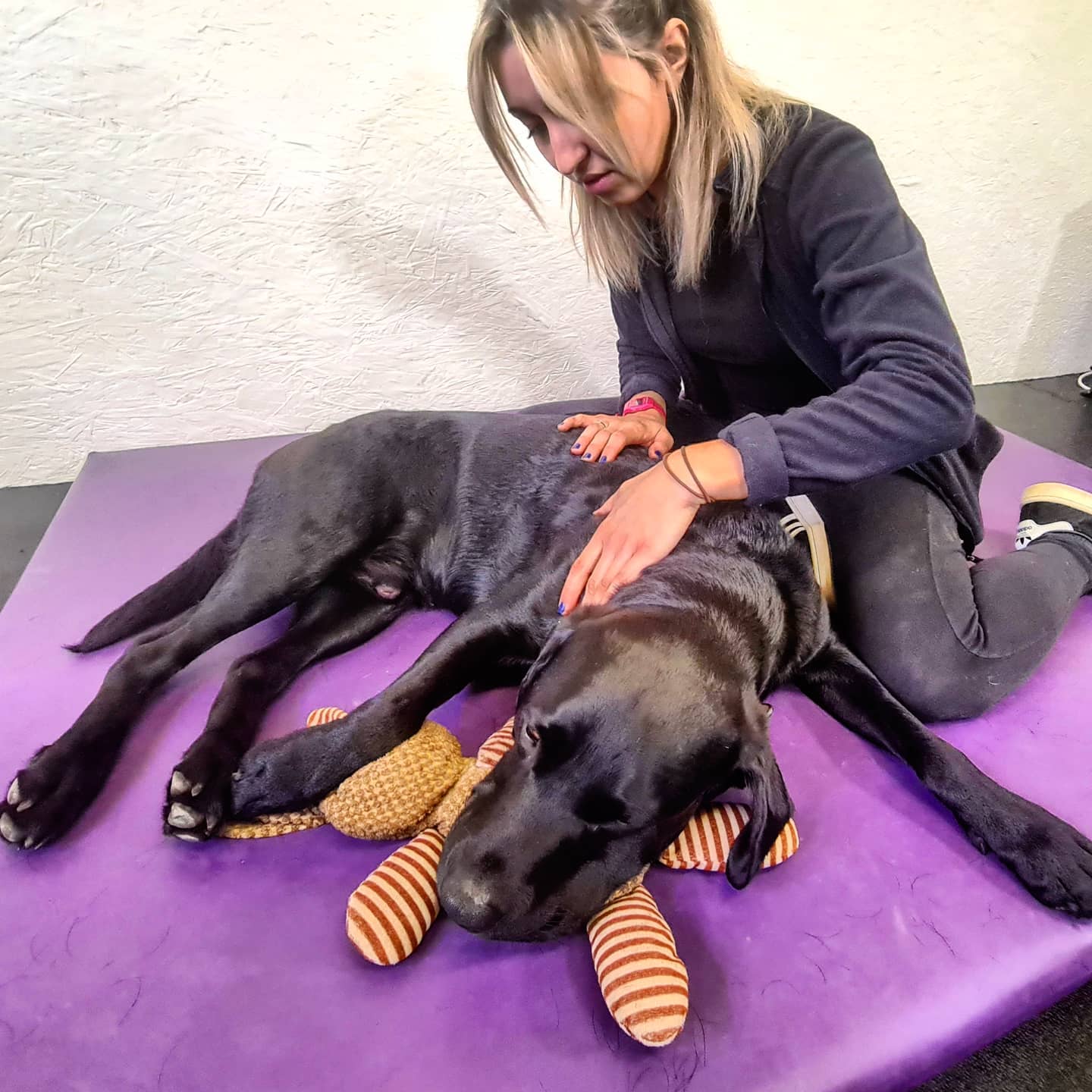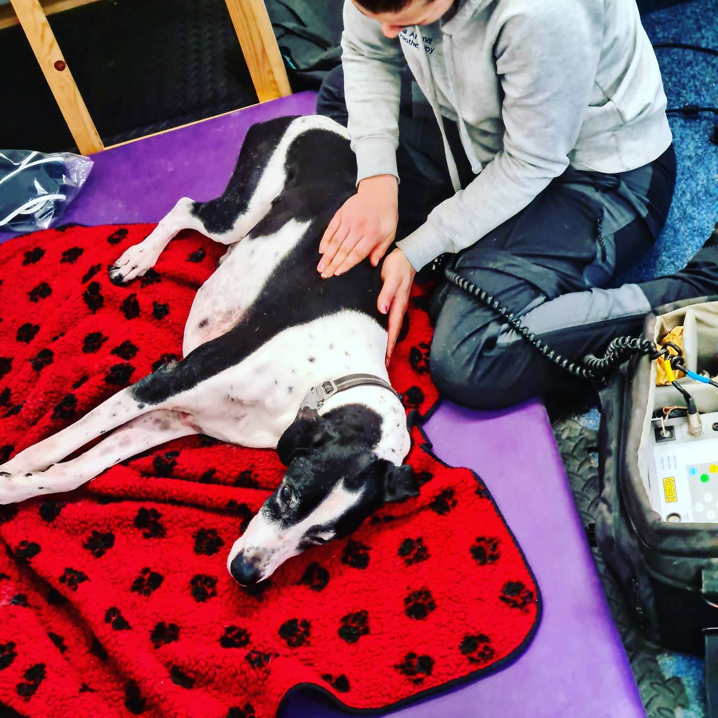
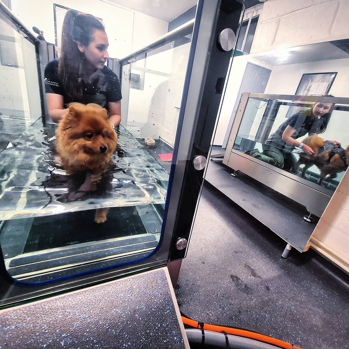
If your dog has recently had surgery, suffered an injury, or is dealing with a chronic condition like arthritis, you may be considering physiotherapy to aid their recovery. One of the most common questions dog owners ask is: How many physiotherapy sessions will my dog need?
After analysing data from over 5,000 rehabilitation bookings, we’ve found that, on average, dogs require 10.13 physiotherapy appointments. The typical rehabilitation cost comes to £519.74. But what do these figures mean for your pet?
What Affects the Number of Physiotherapy Sessions?
Every dog is different, and the number of sessions needed for recovery depends on several factors:
- Type of Injury or Condition
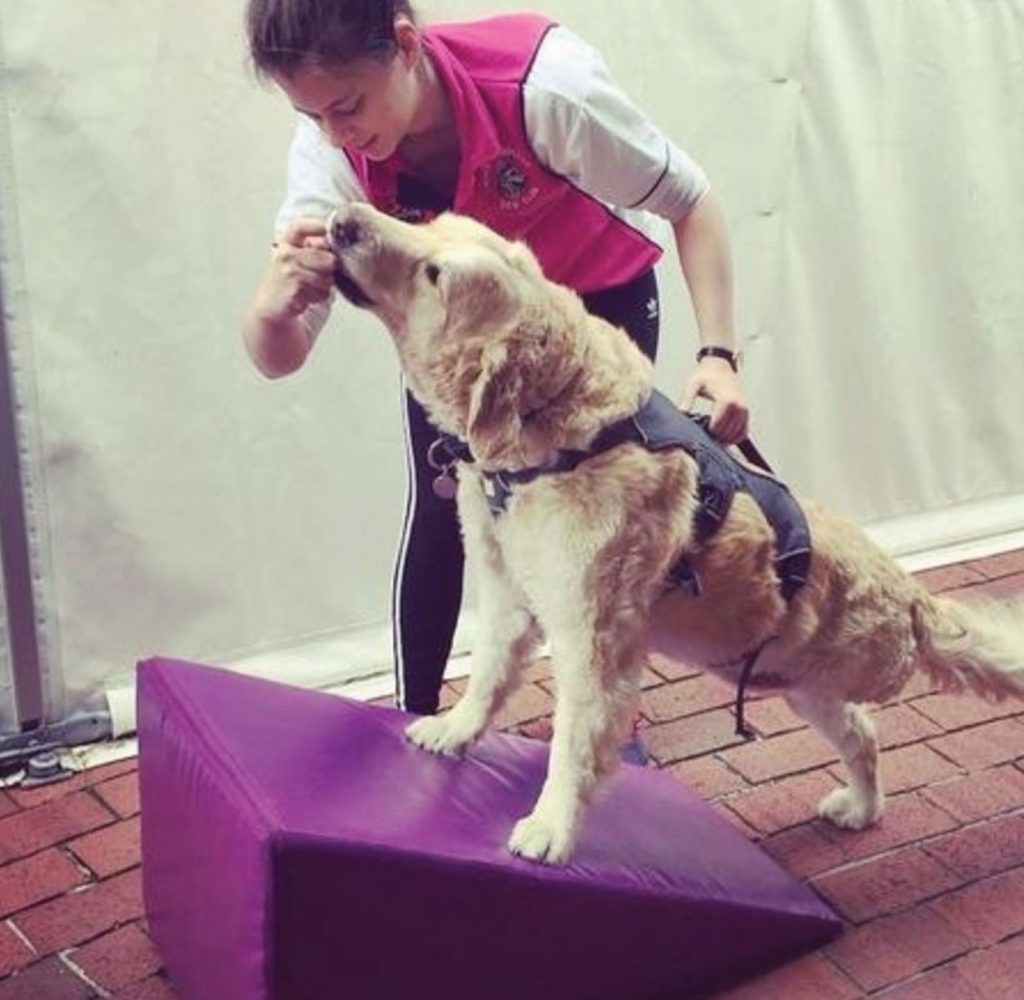
- Post-surgery rehabilitation, such as for cruciate ligament repairs or hip dysplasia, often involves 6 to 12 weeks of physiotherapy, with dogs attending 1 to 3 sessions per week.
- Chronic conditions like arthritis may require ongoing therapy, starting with 1 or 2 sessions per week, which can be adjusted depending on your dog’s progress.
- For acute injuries like sprains, your dog might need 4 to 8 sessions over a few weeks.
- Neurological conditions, such as paralysis or nerve damage, often call for more intensive, long-term physiotherapy, sometimes lasting several months.
- Age and Overall Health
Older dogs or those with underlying health issues may need more sessions due to slower recovery. On the other hand, younger, healthier dogs might show quicker improvement, possibly reducing the number of appointments required. - Response to Treatment
Dogs respond to physiotherapy at different rates. Some may show significant progress after just a few sessions, while others might need a more extended course of treatment to fully recover.
How Much Will You Spend on Canine Physiotherapy?
Alongside the number of sessions, the cost is another key concern for dog owners. Our data reveals that the average spend per dog is £519.74, though this can vary based on factors such as:
- Location: Treatment costs can differ depending on where you live.
- Condition complexity: More complicated cases that need specialised equipment or techniques may increase the overall rehabilitation costs.
- Session frequency: Dogs needing more frequent sessions, especially early in their treatment, may see higher initial costs.
Is Physiotherapy for Dogs Worth the Investment?
With an average of £519.74 spent and around 10.13 sessions required, many pet owners wonder if physiotherapy is worth it. The benefits, however, can be substantial:
- Improved mobility and comfort: For dogs with chronic conditions like arthritis, physiotherapy can significantly improve their ability to move and their overall comfort.
- Faster recovery from surgery or injury: Physiotherapy helps strengthen key muscle groups, improving flexibility and speeding up the healing process, so your dog can return to their normal activities sooner.
- Reduced reliance on medication: Regular physiotherapy can reduce pain and inflammation, potentially lessening the need for long-term use of medications, which may have side effects.
Tailoring a Treatment Plan for Your Dog
While these averages offer a helpful guide, each dog’s treatment plan should be tailored to their specific needs. A veterinary physiotherapist will assess your dog’s condition and recommend a course of therapy based on their individual situation. Regular monitoring and adjustments to the treatment plan will ensure your dog gets the best possible care.
Conclusion
From our analysis, we’ve found that the average dog undergoes 10.13 physiotherapy sessions and the average cost is £519.74. However, these figures are just a starting point. Your dog’s requirements may vary, but the goal remains the same: ensuring they lead a healthy, happy, and pain-free life. Physiotherapy can be an essential part of your dog’s recovery and long-term wellbeing.
If you’re unsure whether physiotherapy is right for your dog, it’s always best to consult your vet. They can advise on the most suitable treatment options for your pet’s individual needs.