
Chronic pain in pets, much like in humans, can significantly impact their quality of life. It often arises from conditions like arthritis, spinal problems, or long-term injuries, and managing it requires a comprehensive, multi-disciplinary approach. Veterinary professionals and physiotherapists work together to offer tailored treatments that focus on reducing pain, improving mobility, and enhancing a pet’s overall well-being.
In this blog, we’ll explore how vets and physios collaborate to create a pain management strategy for pets suffering from chronic pain, and how their combined efforts lead to better outcomes.
1. Assessment: The First Step to Pain Management
The process of managing chronic pain in pets begins with an accurate diagnosis, which typically involves a veterinarian. Vets play a crucial role in identifying the source of pain through a variety of methods, such as physical examinations, diagnostic imaging (like X-rays or MRIs), and sometimes blood tests to rule out underlying diseases.
Once the diagnosis is confirmed, the vet develops a treatment plan that often includes pain medications, anti-inflammatory drugs, and sometimes dietary changes. However, managing chronic pain is more than just about medications; it’s about addressing the underlying mobility issues and restoring function—this is where physiotherapists enter the picture.
2. The Role of Physiotherapists in Pain Relief
Veterinary physiotherapists are specialists in movement and rehabilitation. After a vet’s diagnosis, a physio steps in to assess the pet’s mobility, muscle strength, and joint function. The goal is to alleviate pain by improving the animal’s physical condition, helping them move more comfortably, and reducing the strain on painful joints or injured areas.
Here are some key methods that physios use to manage chronic pain:
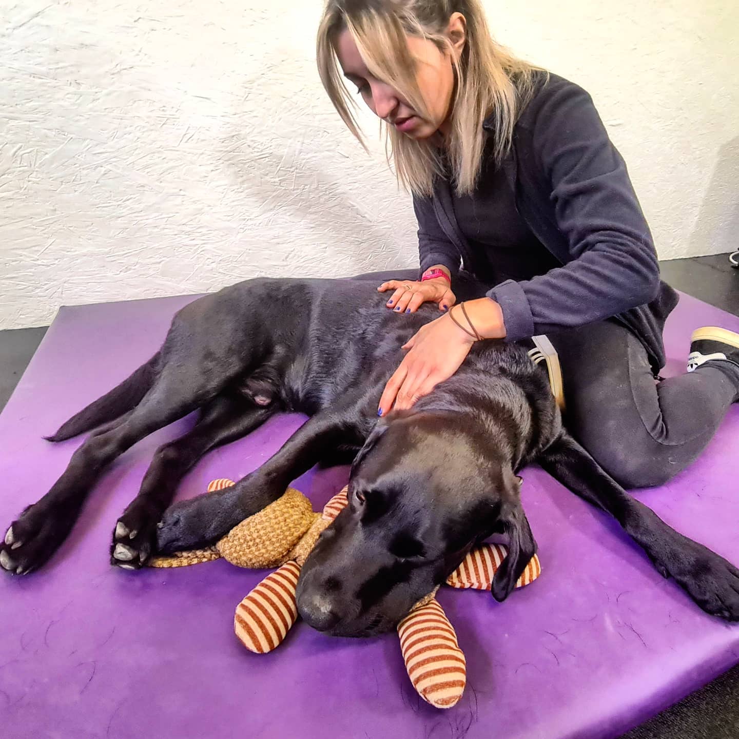
- Manual Therapy: This includes massage, stretching, and manipulation techniques to relieve muscle tension and improve joint mobility.
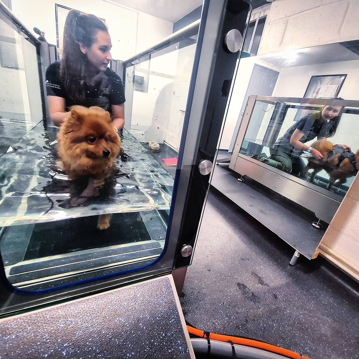
- Hydrotherapy: Water-based exercises, like swimming or underwater treadmill work, allow pets to move with reduced weight-bearing stress on their joints, which helps them exercise without exacerbating pain.
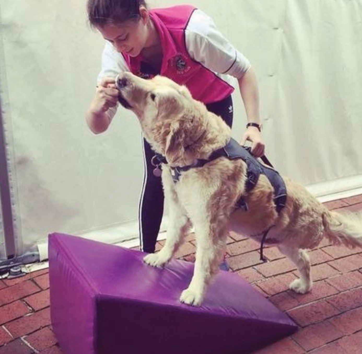
- Therapeutic Exercises: Tailored exercises are designed to strengthen muscles and improve flexibility, supporting the joints and reducing discomfort in daily activities.
- Laser Therapy and Ultrasound: These non-invasive treatments can help reduce inflammation, accelerate healing, and improve circulation, all of which contribute to pain relief.
3. Coordinated Care: Communication Between Vets and Physios
The collaboration between vets and physiotherapists is essential for effective pain management. Regular communication ensures that the treatments are complementary and not contradictory. For example, a vet may prescribe certain medications to reduce inflammation, while a physio focuses on restoring mobility through exercise. Together, they monitor the pet’s progress, adjusting the treatment plan as needed.
A typical treatment strategy may involve regular veterinary check-ups to assess the pain levels and adjust medications, combined with ongoing physiotherapy sessions to improve strength and mobility.
This synergy allows for:
- Personalized Treatment Plans: Every pet responds differently to treatments, and what works for one may not work for another. By working together, vets and physios can modify treatment plans based on real-time feedback, ensuring that the care remains adaptive and effective.
- Long-Term Monitoring: Chronic pain is a condition that requires ongoing management. The combined efforts of vets and physios help track the pet’s progress, whether it’s reducing medication doses or increasing exercise intensity as mobility improves.
4. Preventative Strategies and Lifestyle Changes
Beyond managing current pain, vets and physiotherapists collaborate on preventative strategies to avoid worsening conditions. This could involve:
- Weight Management: Obesity can exacerbate joint pain, particularly in pets with arthritis. Both vets and physios often advise owners on maintaining an ideal weight through a balanced diet and controlled physical activity.
- Environmental Modifications: Physiotherapists might suggest changes at home to make life easier for pets, such as using ramps to avoid stairs or cushioned beds to reduce pressure on painful joints.
- Ongoing Exercise Plans: Physiotherapists provide pet owners with exercises they can do at home, ensuring that pets continue to stay active and maintain muscle strength between professional sessions.
5. Improving Quality of Life
The ultimate goal of veterinary and physiotherapy collaboration is to improve a pet’s quality of life. Pets, just like people, deserve to live without constant pain. By addressing not only the symptoms of chronic pain but also the underlying causes, vets and physios offer a holistic approach to care. They ensure that pets can enjoy a higher degree of mobility, engage with their families, and lead more fulfilling lives despite their chronic conditions.
Conclusion
Chronic pain in pets is complex and often requires more than just medication to manage. The collaboration between vets and physiotherapists brings together the best of both worlds—medical knowledge and physical rehabilitation—to create a comprehensive, customized treatment plan for each pet. By working together, they can effectively manage chronic pain, offering pets a chance to live more active, pain-free lives.
For pet owners, this partnership provides peace of mind, knowing that their pets are receiving well-rounded, expert care that targets both immediate pain relief and long-term well-being.